fbpx

What is Leukemia?

Leukemia is cancer of the white blood cells. White blood cells, which are also called leukocytes, fight against infections and other diseases. In leukemia, the bone marrow produces many abnormal white blood cells, and these abnormal white blood cells enter the bloodstream by filling the bone marrow. Unlike healthy white blood cells, these cannot protect the body from infections. Leukemia is the most common type of cancer amongst children. 

What are the Types of Leukemia? 

There are many different types of leukemia. The most common type in children is Acute Lymphocytic Leukemia, ALL for short. Less common types of childhood leukemia can be listed as Acute Myeloid Leukemia, Chronic Myeloid Leukemia (CML) and Juvenile Myelomonocytic Leukemia. 

What are the Signs and Symptoms of Leukemia? 

Because their white blood cells cannot fight against infections, children with leukemia may get more viral or bacterial infections compared to other children. In addition, anemia or exsanguinity is also often common. Other symptoms of leukemia are: anemia, pale appearance, pain in bones or joints, swollen lymph nodes (swollen glands) in the neck, loss of appetite, weight loss, irresolute fever, the body getting bruised easily, nosebleeds, swelling in the gums.

How is Leukemia Diagnosed? 

The steps followed to find out if a child has leukemia can be summarized as follows; the physician first asks questions about the symptoms. Then, they examine them to check for signs of infection, anemia, unusual bleeding, and swollen lymph nodes. In addition to this examination, some tests may be requested. Depending on the results, other tests such as bone marrow biopsy, spinal needle to obtain lumbar fluid, imaging tests, genetic tests may also be performed. 

How Is Leukemia Treated? 

A pediatric oncologist (a doctor who specializes in childhood cancer) leads the medical team caring for the child with leukemia. The oncologist works together with other specialists, including nurses, social workers, psychologists and surgeons. The main treatment approach in childhood leukemia is chemotherapy. The doses and drugs used may differ depending on the child’s age and the type of leukemia. Other treatment methods such as radiation therapy, targeted drugs, bone marrow transplantation can also be performed.

Some other facts about leukemia:

  1. Your child’s sex can affect your prognosis and how long your treatment will take.
  2. Bone marrow transplantation is not always needed when treating leukemia. This depends on the type of leukemia and the response to treatment.
  3. You should take fever very seriously during leukemia treatment. In most cases, the use of antibiotics should be started immediately. 
  4. You may need to interrupt your child’s vaccinations for a while during the treatment for leukemia.
  5. There may be many long-term side effects associated with leukemia treatment. Depending on the treatment your child receives, post-treatment follow-up is required. 

Grierson-Gopalan Syndrome: Unveiling the Rare Condition

Grierson-Gopalan Syndrome, often abbreviated as GGS, is a medical rarity that has puzzled healthcare professionals and researchers alike due to its extreme scarcity. This complex and enigmatic condition, named after the pioneering doctors who first documented it, Dr. I. D. Grierson and Dr. Ravindra Gopalan, presents a distinctive array of symptoms, primarily affecting craniofacial development, limb structure, and dental health. In this comprehensive guide, we embark on a journey to unravel the mysteries surrounding Grierson-Gopalan Syndrome, exploring its symptoms, genetic factors, diagnostic approaches, and the supportive therapies that can enhance the lives of those affected by this unique condition. Join us in shedding light on this extraordinary medical phenomenon and the hope it holds for future research and treatment.

Exploring Grierson-Gopalan Syndrome

Grierson-Gopalan Syndrome (GGS) remains a medical enigma, with its extreme rarity posing significant challenges for diagnosis and understanding. This section delves deeper into the complexities of GGS, shedding light on its historical context, symptoms, genetic factors, and the intricacies surrounding its diagnosis and treatment. By exploring the depths of this rare condition, we aim to contribute to the growing body of knowledge about GGS and its impact on those who experience it.

Understanding the Rarity

Grierson-Gopalan Syndrome (GGS) stands as a medical enigma due to its extreme rarity. This condition affects only a small number of individuals worldwide, making it a formidable challenge for the medical community. The scarcity of cases has contributed to the intrigue surrounding GGS, prompting further research to uncover its mysteries.

Historical Perspective

The historical journey of Grierson-Gopalan Syndrome is fascinating. It was named after Dr. I. D. Grierson and Dr. Ravindra Gopalan, who first documented the condition in the early 1980s. Since then, researchers and healthcare professionals have been working diligently to understand this unique syndrome better.

Grierson-Gopalan Syndrome Symptoms

Grierson-Gopalan Syndrome (GGS) manifests a unique constellation of symptoms that set it apart as a distinct medical condition. From craniofacial anomalies that shape one’s appearance to limb abnormalities that impact mobility, and dental issues that affect oral health, GGS presents a complex clinical profile. This section delves into the specific symptoms and characteristics that define GGS.

Craniofacial Anomalies

One of the distinctive features of GGS is its impact on craniofacial development. Individuals with GGS often exhibit craniofacial anomalies, which may include facial asymmetry and unique facial features. These characteristics contribute to the diagnostic criteria for GGS.

Limb Abnormalities

GGS can also manifest in limb abnormalities, impacting the structure and function of hands and feet. These limb anomalies can vary in severity, affecting mobility and daily activities.

Dental Issues

Dental problems are another common aspect of GGS. Individuals with this syndrome may experience dental anomalies such as missing teeth or irregular dental development, necessitating specialized dental care.

Causes and Genetic Factors

Understanding Grierson-Gopalan Syndrome (GGS) requires a closer look at the genetic underpinnings that contribute to its development. GGS is primarily attributed to genetic mutations, yet the precise genes involved and the mechanisms behind these mutations remain subjects of ongoing research. Uncovering the genetic factors at play not only enhances our understanding of GGS but also holds the potential to guide future diagnostic and therapeutic advances.

Genetic Mutations

Grierson-Gopalan Syndrome is primarily attributed to genetic mutations. However, the specific genes involved and the mechanisms triggering these mutations remain subjects of ongoing research. Identifying the genetic underpinnings of GGS is essential for improved diagnosis and potential targeted treatments.

Inheritance Patterns

Understanding the inheritance patterns of GGS is crucial for affected individuals and their families. While GGS is a rare condition, knowledge about its hereditary nature can guide genetic counseling and family planning decisions.

Diagnosis and Medical Evaluation

Diagnosing Grierson-Gopalan Syndrome (GGS) presents a unique set of challenges due to its extreme rarity and diverse array of symptoms. Healthcare professionals rely on comprehensive clinical assessments that encompass physical examinations and reviews of medical history to identify characteristic features associated with GGS. Additionally, genetic testing plays a pivotal role in confirming the diagnosis and pinpointing specific genetic mutations linked to the syndrome.

Clinical Assessment

Diagnosing GGS typically involves a comprehensive clinical assessment. Medical professionals rely on physical examinations and reviews of medical history to identify characteristic features and symptoms associated with the syndrome. While GGS may not always present with overt symptoms, its distinct clinical characteristics aid in the diagnostic process.

Genetic Testing

Advancements in genetic testing have significantly impacted the diagnosis of rare conditions like GGS. Genetic tests can pinpoint specific genetic mutations associated with the syndrome, enhancing diagnostic accuracy and facilitating genetic counseling for affected individuals and their families.
Grierson-Gopalan Syndrome Treatment

Multidisciplinary Care

Managing GGS requires a multidisciplinary approach involving specialists in genetics, orthopedics, dentistry, and other relevant fields. Coordinated care ensures that individuals with GGS receive comprehensive treatment addressing their specific needs.

Supportive Therapies

While there is currently no cure for GGS, supportive therapies can improve the quality of life for affected individuals. Physical and occupational therapies may help address limb abnormalities and enhance mobility. Dental interventions can address oral health issues associated with GGS, offering relief and improved dental function.

Is Grierson-Gopalan Syndrome Serious?

Grierson-Gopalan Syndrome is indeed a serious medical condition, primarily due to its impact on craniofacial development, limb structure, and dental health. The severity of the syndrome can vary among individuals, with some experiencing more pronounced symptoms and challenges than others. Early diagnosis and a multidisciplinary approach to care are essential for managing the condition effectively and addressing its potential complications.

Can Grierson-Gopalan Syndrome Be Cured?

As of now, there is no known cure for Grierson-Gopalan Syndrome. However, ongoing research into the genetic underpinnings of the syndrome holds promise for future treatments. While a cure may not yet be within reach, supportive therapies and multidisciplinary care can significantly improve the quality of life for individuals living with GGS.

Grierson Gopalan Syndrome B12

Vitamin B12 deficiency is not directly associated with Grierson-Gopalan Syndrome itself. However, individuals with GGS may require specialized nutritional support, including vitamin B12 supplementation, to address any nutritional deficiencies that may arise due to their unique dietary needs or dental issues associated with the syndrome.

What is Osteoporosis?

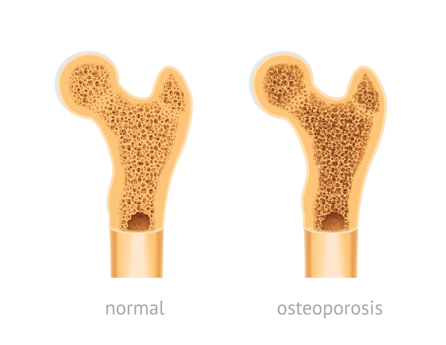
Osteoporosis, a condition that weakens bones and makes them more susceptible to fractures, is a significant concern for many individuals, especially as they age. Often referred to as a “silent disease,” osteoporosis can silently progress without apparent symptoms until a fracture occurs. This comprehensive guide delves into the world of osteoporosis, shedding light on its symptoms, risk factors, treatment options, and the critical role of exercise in bone health. Whether you’re seeking to understand this condition better or looking for guidance on managing it, this informative resource aims to provide you with the knowledge and insights you need to maintain strong and healthy bones.

Understanding Osteoporosis

Osteoporosis, often referred to as a “silent disease,” is a common bone disorder characterized by weakened bones. It’s a condition that tends to progress stealthily, often without any noticeable symptoms until a fracture or break occurs. To grasp the significance of osteoporosis and how to best manage it, it’s crucial to start with the basics.

Bone Composition

Bones are remarkable structures in our bodies. They provide essential support, protect vital organs, and enable movement. To understand osteoporosis, it’s important to know what bones are made of. They consist of minerals, primarily calcium and phosphate, which give them their density and strength. Additionally, bones contain collagen, a protein that provides flexibility.

Bone Remodeling

A dynamic process called bone remodeling is constantly taking place in our bodies. This process involves the breakdown of old bone tissue and the formation of new bone. It’s a finely tuned system that ensures the maintenance of strong and healthy bones. However, in osteoporosis, this balance is disrupted, resulting in the gradual weakening of bone structure.

Osteoporosis Symptoms

Osteoporosis is often a silent condition until a fracture or break occurs. However, there are some common symptoms and signs to be aware of:

Fractures

Fractures, particularly in the hip, spine, and wrist, are hallmark signs of osteoporosis. These fractures can occur with minimal trauma or even during everyday activities. A simple stumble or fall can result in a significant fracture.

Loss of Height

Over time, vertebral fractures can lead to a noticeable loss of height and a stooped posture, which is often referred to as kyphosis. This change in posture can affect one’s overall quality of life.

Back Pain

Chronic back pain, especially in the lower back, can be an indication of osteoporosis. This pain may result from vertebral fractures or the compression of spinal nerves.

Osteoporosis Risk Factors

Certain factors can increase an individual’s risk of developing osteoporosis:

Age and Gender

Osteoporosis is more commonly seen in older individuals, and women, particularly after menopause, are at a higher risk. This is because hormonal changes associated with aging can affect bone density.

Family History

A family history of osteoporosis can elevate your risk. Genetic factors can play a significant role in bone health.

Hormonal Changes

Conditions or treatments that impact hormone levels, such as low estrogen in women or low testosterone in men, can contribute to bone loss.

Diet and Nutrition

Inadequate intake of essential nutrients, particularly calcium and vitamin D, can weaken bones. These nutrients are vital for maintaining bone density.

Lifestyle Factors

Certain lifestyle choices can increase the risk of osteoporosis. These include a sedentary lifestyle, smoking, excessive alcohol consumption, and maintaining a low body weight.

Diagnosis and Bone Density Testing

Early diagnosis is key to effectively managing osteoporosis. Bone density testing, often conducted through Dual-Energy X-ray Absorptiometry (DXA) scans, is a crucial tool in assessing bone health. These tests measure bone mineral density and help determine the presence and severity of osteoporosis.

Osteoporosis Treatment

When it comes to treating osteoporosis, several options are available:

Medications

Various medications are prescribed to slow down bone loss and reduce the risk of fractures. Commonly prescribed osteoporosis medications include bisphosphonates, denosumab, and hormone therapy. The choice of medication depends on individual factors such as age, sex, and the severity of osteoporosis.

Lifestyle Changes

Lifestyle modifications can significantly impact bone health. Implementing a well-balanced diet rich in calcium and vitamin D is crucial. Weight-bearing and resistance exercises, such as walking or strength training, can help strengthen bones. Quitting smoking and reducing alcohol consumption are also recommended.

Fall Prevention

Taking precautions to prevent falls is essential, especially for individuals with osteoporosis. Simple measures like improving home safety by removing tripping hazards and installing handrails can go a long way in preventing accidents.

Living with Osteoporosis

Osteoporosis is a manageable condition, and individuals can lead fulfilling lives with the right care and support:

Support and Education

Joining support groups for individuals with osteoporosis can provide valuable emotional and practical support. Staying informed about the condition and its management options empowers individuals to take an active role in their healthcare.

Regular Check-Ups

Routine follow-up appointments with healthcare providers are crucial for monitoring bone health. These appointments include bone density testing and discussions about medication effectiveness and any necessary adjustments.

What is the Best and Safest Treatment for Osteoporosis?

The choice of osteoporosis treatment varies from person to person and should be determined by a healthcare professional based on an individual’s specific needs and risk factors. What may be the best and safest treatment for one person might not be the same for another. Therefore, it’s essential to consult with a healthcare provider to develop a personalized treatment plan.

Exercise for Osteoporosis

Exercise is a vital component of osteoporosis management. Weight-bearing and resistance exercises can help improve bone density and strength. However, it’s important to engage in exercise under the guidance of a healthcare provider or a qualified fitness professional to ensure safety and effectiveness.

Acoustic Neuroma: Symptoms, Treatment, and Life After Surgery

Acoustic neuroma, also known as vestibular schwannoma, is a non-cancerous tumor that affects the hearing and balance functions. This condition can lead to symptoms like hearing loss, tinnitus, and balance issues, making it essential to understand its causes, treatment options, and life after surgery. In this guide, we’ll explore the world of acoustic neuroma, offering insights into its diagnosis, treatment, and the journey to recovery.

What is an Acoustic Neuroma?

Acoustic neuroma, also known as vestibular schwannoma, is a non-cancerous tumor that typically grows on the vestibular nerve, which connects the inner ear to the brain. While these tumors are usually benign, they can cause a range of symptoms that affect hearing and balance. Let’s delve into the details of acoustic neuromas, including their symptoms, treatment options, and what life may look like before and after surgery.

Symptoms of Acoustic Neuroma

The symptoms of acoustic neuromas can vary from person to person, but there are some common indicators to watch out for:

Hearing Loss

One of the most prevalent signs of acoustic neuroma is hearing loss. This condition often begins with gradual or sudden hearing impairment in one ear, affecting your ability to communicate effectively.

Tinnitus

Many individuals with acoustic neuromas experience persistent ringing or buzzing in the affected ear, a condition known as tinnitus. This symptom can be distracting and disruptive to daily life.

Balance Issues

Acoustic neuromas can lead to balance and coordination problems, resulting in unsteadiness and difficulty maintaining equilibrium. This symptom can affect your mobility and quality of life.

Ear Fullness

Some patients report a sensation of fullness or pressure in the ear affected by the tumor. This discomfort can be unsettling and bothersome.

Headaches

While not as common as other symptoms, occasional headaches, usually on the side of the affected ear, can occur in individuals with acoustic neuromas.

Facial Numbness

In rare cases, acoustic neuromas may cause numbness or weakness on one side of the face. This symptom warrants immediate medical attention.

Causes of Acoustic Neuroma

The precise cause of acoustic neuromas isn’t always clear, but several factors may contribute to their development:

  • Genetic Mutation (NF2): Some cases of acoustic neuroma are associated with a genetic mutation called NF2. Individuals with a family history of this condition may be at a higher risk.
  • Radiation Exposure: Exposure to ionizing radiation, such as radiation therapy for previous head and neck conditions, is a potential risk factor.
  • Rare Genetic Disorders: Certain rare genetic disorders, though uncommon, can increase the likelihood of developing acoustic neuromas.

Diagnosis of Acoustic Neuroma

Diagnosing acoustic neuroma involves a series of evaluations to confirm the presence and extent of the tumor:

Hearing Tests

Audiometric tests are commonly employed to assess hearing loss and determine its severity. This initial evaluation helps healthcare providers gauge the extent of the issue.

Imaging

Magnetic Resonance Imaging (MRI) scans are essential for visualizing the size and location of the acoustic neuroma. This imaging technique provides critical information for treatment planning.

Monitoring

In cases where the tumor is small or not causing significant symptoms, doctors may recommend regular monitoring to observe any changes in size or symptoms over time.

Treatment Options for Acoustic Neuroma

The choice of treatment for acoustic neuroma depends on various factors, including the size of the tumor and the patient’s overall health:

Observation (Watch and Wait)

For small, slow-growing tumors, a strategy of “watchful waiting” may be appropriate. This approach involves monitoring the tumor’s progression and intervening only if it starts to grow or cause more severe symptoms.

Surgery

Surgery is a common treatment option for acoustic neuromas, and several surgical approaches can be employed:

Translabyrinthine Surgery

This surgical procedure involves removing the tumor through the ear canal. It is typically suitable for larger tumors and offers a direct path to the tumor.

Middle Fossa Surgery

Middle fossa surgery targets tumors located near the inner ear. It aims to remove the tumor while preserving hearing and facial nerve function.

Retrosigmoid Surgery

Retrosigmoid surgery accesses the tumor from behind the ear. This approach is often chosen when hearing preservation is a primary concern.

Radiation Therapy

Radiation therapy, including techniques like stereotactic radiosurgery, may be recommended for patients who cannot undergo surgery or prefer non-invasive treatments. This approach uses targeted radiation to shrink or control the tumor’s growth.

Life Expectancy After Acoustic Neuroma Surgery

Undergoing surgery for an acoustic neuroma can be a life-changing experience. It’s essential to understand what to expect both before and after the procedure:

Before Acoustic Neuroma Surgery

Before surgery, you will undergo a thorough evaluation to assess your overall health and the specifics of your tumor. Your medical team will discuss the surgical approach and potential risks. It’s crucial to have realistic expectations regarding hearing preservation and facial nerve function.

After Acoustic Neuroma Surgery

Life expectancy after acoustic neuroma surgery varies from person to person, but there are some common considerations:

  • Recovery and Rehabilitation: Rehabilitation, including physical therapy, may be necessary to regain balance and adapt to changes in hearing.
  • Coping with Hearing Loss: If hearing preservation is not possible, you may explore options such as hearing aids or learning sign language to enhance communication.
  • Support Groups: Joining support groups for individuals facing similar challenges can provide emotional and practical support during your recovery.

Prognosis and Follow-Up

Regular follow-up appointments are essential after acoustic neuroma surgery to monitor your progress and ensure that the tumor does not return. The prognosis for individuals with acoustic neuromas is generally positive when detected early and managed appropriately.

Potential Complications

While complications are rare, it’s important to be aware of possible issues, including facial nerve damage, further hearing loss, and ongoing balance problems. Prompt medical attention can address these concerns if they arise.

What is Melatonin?

Melatonin, often referred to as the body’s internal timekeeper, plays a very important role in your sleep-wake cycle aka. circadian rhythm. This hormone, primarily released at night, not only influences sleep but also impacts hormone secretion, body temperature, and mood.

Understanding Melatonin

To truly appreciate the significance of melatonin, we must learn more about this remarkable hormone. That’s why in this section, we’ll discover everything abot melatonin from how it is synthesized in the body to its effects.

What is Melatonin Exactly?

Melatonin is a naturally occurring hormone produced by the pineal gland in the brain. It actually is released in response to darkness, signaling to the body that it’s time to prepare for sleep. Which means the production of melatonin is influenced by external factors, primarily light and darkness.

The Circadian Rhythm Connection

The circadian rhythm, which controls sleep patterns, hormone release, body temperature and many other bodily fuctions, has a strong relation with the melatonin hormone. As day passes into night and darkness falls, melatonin levels begin to rise. This cause us to feel sleepy and ready for sleep. So the melatonin secrecation is linked with the abscence of daylight exposure. This natural process helps us maintain a healthy sleep schedule and stay synchronized with the day-night cycle.

Uses of Melatonin

Melatonin, as a natural product of your body can also be used as a supplement. Many individuals struggling sleep problems use melatonin supplements to recover. So in this part we’ll examine the external uses of melatonin.

What is Melatonin Used For?

Melatonin supplements are commonly used to address sleep-related issues. Individuals experiencing sleep disorders, such as insomnia, often turn to melatonin to help regulate their sleep patterns. But it’s not the only use of melatonin as a supplement. For example people who experience jetlags or shift workers can also use it to organize their sleep patterns.

Beyond Sleep: Melatonin’s Surprising Benefits

Yes, melatonin is first recognized as a sleep hormone and we use it mainly on this purpose. However there are some

Melatonin Benefits and Side Effects

The advantages of melatonin extend beyond its primary function, but it’s essential to understand both its benefits and potential drawbacks. In this section, we’ll delve into the potential health benefits of melatonin, such as its antioxidant properties, while also addressing safety considerations and potential side effects.

What is Melatonin Good For?

One of the key benefits of melatonin is its role as an antioxidant. It helps protect cells from oxidative stress, which is linked to various chronic diseases. Melatonin’s antioxidant properties may contribute to its potential for reducing the risk of certain health issues.

Melatonin Side Effects and Safety

While melatonin is generally considered safe for short-term use, it’s essential to be aware of potential side effects. These side effects may include drowsiness, dizziness, and changes in blood pressure. It’s crucial to consult with a healthcare professional before starting melatonin supplementation, especially if you have underlying health conditions or are taking other medications. Proper dosage and timing are also important to avoid unwanted side effects.

Natural Sources and Supplements

While melatonin is naturally produced in the body, we can also influence its levels through dietary choices. Certain foods contain melatonin or compounds that promote its production. We’ll begin by exploring these natural sources and then dive into the world of melatonin supplements, discussing variations and considerations for selecting the right one.

Natural Sources of Melatonin

Melatonin can also be obtained through dietary choices. Some foods naturally contain melatonin or compounds that promote its production. Cherries, nuts, and seeds, for example, are known to contain melatonin precursors. Including these foods in your diet may help support healthy melatonin levels.

Melatonin Supplements: A Closer Look

For those who have difficulty obtaining enough melatonin from natural sources, supplements are readily available. Melatonin supplements come in various forms, including pills, capsules, and even gummies. It’s essential to choose a reputable brand and follow dosage recommendations carefully.

Tips for Using Melatonin

To make the most of melatonin’s benefits while minimizing potential side effects, it’s essential to use it wisely. This section offers guidance on determining the appropriate melatonin dosage based on individual needs and outlines best practices for effectively and safely using melatonin supplements.

Dosage Recommendations

The right melatonin dosage varies from person to person, influenced by factors such as age, sleep disorder severity, and individual sensitivity. We’ll provide recommendations to help you navigate this aspect wisely, ensuring you get the desired results without unwanted side effects.

Best Practices for Taking Melatonin

Using melatonin supplements effectively involves more than just the right dosage. We’ll offer a set of best practices to enhance your melatonin experience, including tips on establishing a consistent sleep schedule, creating a sleep-conducive environment, and avoiding sleep disruptors like caffeine and alcohol.

Dental Cyst

Dental or odontogenic cysts are fluid-filled sacs within the jawbone or around tooth roots. While most of them are benign, the consequences of untreated dental cysts can be far-reaching, affecting adjacent teeth, bone structure, and overall oral well-being. This comprehensive guide aims to shed light on the various types of dental cysts, their symptoms, potential risks, and the significance of timely diagnosis and treatment. Join us on this educational journey to raise awareness and gain valuable insights into managing dental cysts effectively.

What is a Dental Cyst?

A dental cyst, also referred to as an odontogenic cyst, is a sac packed with fluid that forms inside the jawbone or around tooth roots. These cysts originate from tissues associated with tooth development. Certain cells in the tissues of the mouth may become caught when teeth develop and expand, resulting in fluid collection and cyst formation. The connection between dental cysts and tooth development highlights the developmental nature of these cysts. Unlike other oral conditions, dental cysts specifically emerge during tooth formation. Although most dental cysts are benign, their growth can exert pressure on nearby structures, causing pain, swelling, and potential damage to teeth and bone.

Don’t let Dental Cyst steal your smile. Discover empathetic care and advanced treatments at Liv Hospital in Turkey. Visit us to learn how we can help restore your oral health.

Are Dental Cysts Dangerous?

Dental cysts, in general, tend to be benign, meaning they are not cancerous. This in no way minimizes the importance of their existence in the mouth. Although many dental cysts don’t bring about instant adverse effects, they can progress to complications if left untreated. Dental cysts can exert pressure on neighboring teeth and the jawbone as they grow, potentially causing pain, swelling, tooth displacement, or bone damage. In severe cases, untreated cysts can lead to infections that may spread to other parts of the face and neck. Regular dental check-ups and X-rays are crucial in identifying dental cysts early, allowing for prompt intervention and reducing the risk of further complications.

Your oral health matters to us. Explore Liv Hospital’s expertise in treating Dental Cysts in Istanbul, and let us provide the understanding and care you deserve.


Dental Cyst on Gum

Dental cysts on the gums can develop due to various factors. These cysts often arise from the remnants of dental tissue left behind during tooth development. These fragments can form a cyst when trapped in the gum tissue. Additionally, cysts may develop around the neck of an impacted tooth, where the tooth attempts to erupt but becomes obstructed, leading to fluid accumulation and cyst formation. Identifying symptoms that may indicate the presence of a gum cyst is crucial. These may include localized swelling, tenderness, a small lump or bump on the gum, and discomfort while chewing or speaking.

Dental Cyst Symptoms

Recognizing the symptoms associated with dental cysts is crucial for early detection and timely intervention. Common symptoms of dental cysts include persistent pain or discomfort in the affected area, localized swelling, tenderness to touch, and a noticeable lump or bump on the gums or jaw. Dental cysts associated with non-vital (dead) teeth may cause no pain initially but can lead to swelling and inflammation over time. On the other hand, cysts around impacted teeth might cause visible swelling and difficulty in fully opening the mouth. Regardless of the specific symptoms, prompt recognition is essential to prevent potential complications and ensure effective treatment.

At Liv Hospital, we listen to your concerns and design personalized treatment plans. Contact us in Turkey for information and guidance on your Dental Cyst treatment journey.


Dental Cyst Types

Dental cysts encompass various types, each with distinct characteristics and developmental origins. Radicular cysts, also known as periapical cysts, form at the root tip of a non-vital tooth, often due to an infected pulp. Dentigerous cysts, or follicular cysts, develop around the crown of an unerupted tooth, resulting from fluid accumulation between the tooth and its dental follicle. Odontogenic keratocyst (OKCs) are less common but possess a higher potential for recurrence, affecting the jawbone and displaying aggressive growth. Dental lamina cysts, also called gingival cysts of the newborn, occur in infants due to epithelial remnants from tooth development.

Dealing with Dental cysts may raise questions and concerns, and that’s completely normal. Trust Liv Hospital’s compassionate experts in Istanbul to address your worries and provide the support you need.

Dental Lamina Cysts

Dental lamina cysts, also known as gingival cysts of the newborn, are intriguing anomalies that occur in infants during tooth development. These cysts originate from epithelial remnants left behind by the dental lamina, a structure crucial for tooth formation in the early stages of fetal development. Dental lamina cysts often manifest as small, painless bumps on the gums of newborns. While their appearance may cause concern to parents, it’s important to note that these cysts are generally harmless and self-limiting. Dental lamina cysts usually undergo spontaneous resolution without intervention or treatment.

Dental Cyst Treatment

Treating dental cysts involves various approaches, depending on the cyst’s type, size, and location. Observation and monitoring are often employed for small, asymptomatic cysts that do not pose immediate risks. In cases of larger cysts causing discomfort or swelling, drainage, and aspiration may be performed to relieve pressure temporarily. Enucleation, a surgical procedure, is commonly used to remove the cyst and surrounding tissues altogether. Marsupialization creates an opening in the cyst, allowing continuous drainage to promote healing.

Tooth extraction may be necessary for impacted or severely damaged teeth related to the cyst. Medications like antibiotics can aid in managing infections. Each treatment option serves a specific purpose, and the dentist or oral surgeon determines the most suitable approach based on individual circumstances to ensure effective management of dental cysts.

Liv Hospital – Shaping Healthy Smiles!

Understanding dental cysts and their impact on oral health is vital for maintaining a healthy and pain-free smile. The benign nature of most dental cysts should not be taken lightly, as untreated cysts can lead to complications affecting teeth and surrounding tissues. Regular dental check-ups play a crucial role in early detection and timely intervention, preventing potential problems down the road. If you experience any dental concerns or suspect the presence of a dental cyst, don’t hesitate to seek professional advice.

For those seeking quality healthcare, Liv Hospital, with its leading international vision, stands ready to shape the future of your health in Turkey 2023.

Laryngopharyngeal Reflux

Laryngopharyngeal Reflux (LPR) is a condition often referred to as silent reflux due to its subtle yet significant impact on the upper airway. Raising awareness about LPR is crucial, as its symptoms can be mistaken for other throat conditions, leading to delayed diagnosis and treatment. Throughout this blog, we will delve into the definition and connection between LPR and silent reflux, emphasize the importance of early recognition and explore critical topics encompassing symptoms, treatment options, diet recommendations, natural remedies, and long-term management.

So, let’s uncover the mysteries of LPR and empower ourselves with knowledge for a healthier throat and well-being.

What is Laryngopharyngeal Reflux?

When acid from the gut and digestive enzymes run back toward the larynx and pharynx, producing discomfort and swelling, this condition is known as laryngopharyngeal reflux or LPR. Unlike gastroesophageal reflux disease (GERD), LPR primarily affects the upper airway and doesn’t typically cause heartburn. Chronic hoarseness, coughing, inflammation of the throat, and an obstruction in the throat are some of its symptoms. LPR occurs when the lower esophageal sphincter (LES) fails to close properly, allowing stomach contents to reflux upwards. Contributing factors include diet, lifestyle habits, and certain medications. If left untreated, LPR’s impact on the upper airway can lead to persistent discomfort and potentially damage the vocal cords, underscoring the importance of early recognition and management.

Trust your voice and throat care to Liv Hospital, a leading medical center in Turkey. Our experienced specialists are dedicated to helping you overcome Laryngopharyngeal Reflux.

Recognizing Laryngopharyngeal Reflux Symptoms

Recognizing the silent reflux symptoms is crucial for early diagnosis and effective management. Common symptoms include chronic hoarseness or a rough voice, which may persist despite rest and hydration. Persistent coughing and frequent throat clearing are also prevalent, along with throat irritation and the sensation of a lump in the throat (Globus sensation). LPR can also lead to excessive throat mucus and post nasal drip, contributing to ongoing discomfort. It’s essential to be aware of potential atypical symptoms, such as unexplained ear pain, recurrent sore throat, and mild asthma-like symptoms.

Seeking medical evaluation promptly is vital, as untreated LPR can cause further complications, including vocal cord damage. Individuals can experience relief and safeguard their upper airway health with timely recognition and appropriate care. Restore your voice’s clarity and strength. Discover Liv Hospital’s state-of-the-art treatments for Laryngopharyngeal Reflux in Istanbul and take the first step toward vocal wellness.

Laryngopharyngeal Reflux Treatment

Effective treatment for Laryngopharyngeal Reflux (LPR) involves a multi-faceted approach to alleviate symptoms and promote healing. Lifestyle modifications play a pivotal role, including elevating the head of the bed to prevent nighttime reflux and adopting dietary changes to avoid trigger foods like acidic and fatty items. Weight management and quitting smoking are essential for reducing reflux risk. In the realm of medical treatments, Proton Pump Inhibitors (PPIs) are prescribed to decrease acid production, while H2 blockers mitigate stomach acid effects. In some cases, prokinetics may improve digestive system movement and manage reflux.

When non-surgical procedures fail, an operation may be explored. The lower esophageal sphincter is reinforced by fundoplication, a surgical operation in which the upper stomach coils over the lower esophagus. A personalized treatment plan combining these strategies ensures better outcomes in managing LPR and fostering overall well-being.

Ready to speak freely again? Explore Liv Hospital’s expertise in managing Laryngopharyngeal Reflux and regain control of your voice and throat health. Visit us in Turkey to find out more.

Laryngopharyngeal Reflux Diet: Foods to Avoid and Foods to Include

Maintaining a Laryngopharyngeal Reflux (LPR) diet is crucial to alleviate symptoms and prevent reflux flare-ups. Identifying acidic, spicy, and fatty foods as common triggers is essential, as they can exacerbate reflux and irritation. Opting for a well-balanced diet with various nutrient-rich foods promotes overall health and helps manage LPR effectively. Include reflux-friendly items like lean meats, healthy grains, fruits, and vegetables. Choose non-acidic fruits such as bananas and melons and lean meats such as fish and skinless fowl. Opt for whole grains like oatmeal and quinoa, and incorporate non-citrus vegetables. Additionally, water is the best beverage choice, but herbal teas and non-citrus juices can also be consumed in moderation. Being mindful of your diet empowers you to take control of your LPR symptoms and supports a healthier lifestyle.

Don’t let silent reflux disrupt your daily life. Our caring team at Liv Hospital is here to provide comprehensive care and effective solutions in Istanbul. Find out how we can help you feel better now.

How To Treat Laryngopharyngeal Reflux Treatment Naturally?

Natural remedies and home treatments can complement medical interventions in effectively managing Laryngopharyngeal Reflux (LPR). Emphasizing the potential benefits of natural substances like ginger, slippery elm, and aloe vera is essential, as they possess anti-inflammatory properties that can soothe irritated tissues. Incorporating these ingredients into teas or smoothies can be soothing and aid in symptom relief. Reducing stress levels helps prevent reflux triggers and fosters overall well-being. By embracing these natural remedies and incorporating lifestyle changes, individuals can enhance their journey toward managing LPR more holistically.

Prevention and Long-Term Management

Prevention and long-term management are essential in effectively managing Laryngopharyngeal Reflux (LPR) and promoting throat health. Adopting a reflux-friendly lifestyle, which includes eating a nutritious diet, minimizing trigger meals, and dealing with stress, is critical for preventing LPR flare-ups. Elevating the head of the bed while sleeping can also aid in the reduction of nocturnal reflux. Regular medical follow-ups are essential for monitoring progress and adjusting treatment plans. Emphasizing self-care and attentively monitoring symptoms empowers individuals to take control of their condition and promptly address any changes or concerns. By adopting these proactive measures, individuals can optimize their journey toward lasting relief and long-term well-being.

Liv Hospital –  Leading The Way In LPR Care

understanding Laryngopharyngeal Reflux (LPR) and its impact on the upper airway empowers us to take charge of our throat health. Recognizing the symptoms, seeking an early diagnosis, and embracing a well-rounded treatment approach can lead to effective management and relief. Remember, lifestyle modifications, medical treatments, and natural remedies work hand in hand to ease discomfort and promote healing. At Liv Hospital in Turkey, our commitment to leading international vision drives us to provide our patients with the highest standards of care in Turkey 2023. Experience the future of health with our seven hospitals in Turkey, where compassionate professionals strive to shape a healthier world, one patient at a time. Together, we can face LPR with hope and empowerment, embracing a well-being journey and renewed vocal vitality.

Jock Itch

Jock itch, a prevalent fungal infection, targets the delicate skin of the groin area and inner thighs, causing discomfort and irritation for countless individuals. Despite its common occurrence, many people remain unaware of the insidious nature of this condition, potentially delaying essential treatment. In this informative blog, we delve into the world of jock itch, uncovering its causes, symptoms, and effective treatment options. By shedding light on this often-overlooked topic, we aim to raise awareness about the significance of recognizing the early signs of jock itch. Armed with this knowledge, readers can take proactive measures to tackle the infection promptly, mitigating its impact and restoring comfort and confidence to their daily lives.

What is Jock Itch?

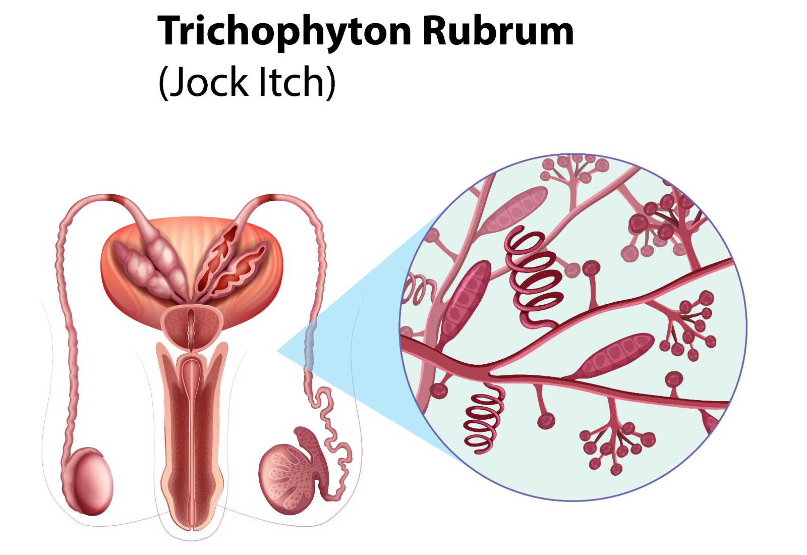
Jock itch, scientifically termed tinea cruris, is a prevalent fungal infection that affects the sensitive skin in the groin and inner thigh regions. The condition presents as a red, itchy rash with a clearly defined border, often forming circular or oval shapes. Dermatophytes, the fungi responsible for athlete’s foot and ringworm, are the culprits behind this uncomfortable infection. These microorganisms thrive in warm and moist environments, finding the groin area to be an ideal breeding ground for their growth and dissemination. As a result, individuals who experience excessive sweating, wear tight clothing, or have poor hygiene practices are at higher risk of developing jock itch.

Don’t let jock itch affect your comfort and confidence. Discover empathetic care and effective treatments at Liv Hospital in Turkey.

Jock Itch Symptoms

Jock itch manifests with a range of distressing symptoms, each contributing to the discomfort experienced by those affected. The most common symptoms include noticeable redness and persistent itching in the groin area and inner thighs. A characteristic rash, often circular or oval in shape, may develop, accompanied by flaky, peeling skin. Individuals may experience a bothersome burning sensation, exacerbating the already irksome condition.

We understand the discomfort and frustration jock itch can cause. Let Liv Hospital’s compassionate experts guide you towards a path of healing and well-being. Find out more by visiting us in Istanbul.

Is Jock Itch Contagious?

Jock itch, though uncomfortable, is not only a concern for the affected individual but also poses a risk of contagion to others. This fungal illness can be transmitted by touching directly with the affected individual’s skin or by swapping personal belongings such as towels, clothes, or sports equipment. To prevent its transmission, simple yet effective measures should be taken. Firstly, avoiding direct contact with infected skin is crucial. Refraining from sharing personal items will minimize the risk of acquiring the infection from others. Maintaining good personal hygiene, especially in the groin area, is essential in preventing jock itch from spreading.

Your well-being is our priority. Explore Liv Hospital’s expertise in treating jock itch, and allow us to provide the understanding and care you deserve in Turkey.

Female Jock Itch

A common misconception surrounding jock itch is that it solely afflicts males. It is essential to debunk this myth and shed light on the fact that females can also develop jock itch. This fungal infection knows no gender bias and can affect women as well. The symptoms experienced by females with jock itch are akin to those in males, such as redness, itching, rash, and discomfort in the groin area and inner thighs. The treatment approaches are similar for both genders, involving the use of antifungal creams and adhering to proper hygiene practices. By understanding this reality, women can be better prepared to recognize the symptoms early and seek timely treatment, enabling them to manage jock itch effectively and regain their comfort and well-being.

Dealing with jock itching may raise questions and concerns, and that’s completely normal. Trust Liv Hospital’s empathetic experts to address your worries and provide the support you need in Istanbul.

How Long Does Jock Itch Last

Several variables can influence the severity of jock itch. If left untreated, the infection can persist for weeks or even months. With timely intervention, the symptoms can be alleviated more quickly. Adherence to treatment is paramount in managing jock itch effectively. Antifungal creams, lotions, or sprays should be applied as directed by a healthcare professional, even if the symptoms start to improve. Incomplete treatment can lead to a resurgence of the infection, prolonging its duration and making it more challenging to control. To ensure a successful outcome, it is vital to complete the full course of antifungal treatment, allowing the body to eradicate the fungus completely and hasten the healing process.

Jock Itch Healing Stages

Jock itch healing progresses through several distinct stages, each marking a step towards recovery. Initially, after commencing antifungal treatment, individuals often experience immediate relief from the relentless itching and discomfort. As the treatment takes effect, the redness and itching gradually diminish, bringing further relief to the affected area. During this phase, the skin’s appearance may begin to improve, with reduced inflammation and flakiness. With consistent adherence to the treatment regimen, the skin restoration process gains momentum.

Jock Itch Treatment

Over-the-counter antifungal creams, lotions, or sprays containing active substances such as clotrimazole, miconazole, terbinafine, or ketoconazole are commonly used to treat jock itch. These medications effectively target the underlying fungal infection and provide relief from symptoms. To ensure successful outcomes, it is crucial to follow proper application guidelines and complete the recommended duration of treatment, even if the symptoms improve. In cases where over-the-counter treatments do not yield desired results, stronger prescription medications may be prescribed by a healthcare professional. Alongside medication, self-care plays a vital role in managing jock itch. Maintaining good personal hygiene, particularly in the groin area, helps prevent further infection.

Prevention of Jock Itch

Preventing jock itch is crucial to avoid the discomfort and inconvenience associated with this fungal infection. Practical tips can be employed to reduce the risk of its occurrence. It is critical to keep the area around your groin neat and dry, particularly following exercise or perspiration. Regularly washing with mild soap and patting dry with a clean towel aids in inhibiting fungal growth. Incorporating antifungal powders in daily hygiene routines helps absorb excess moisture and deters fungal proliferation. Good personal hygiene practices extend beyond cleanliness; refraining from sharing personal items like towels, clothing, and sports equipment is essential to prevent spreading the infection to others or acquiring it from someone else.

Liv Hospital – Compassionate Care, Global Expertise

If you suspect that you may be experiencing jock itch or find that the infection persists despite self-treatment, do not hesitate to seek medical advice. Take charge of your health and well-being by visiting Liv Hospital, a leading institution with a vision of international perfection in healthcare. With seven hospitals across Turkey, Liv Hospital is dedicated to shaping the future of health, ensuring you receive your care in Turkey 2023. Remember, your well-being is of utmost importance, so take the first step towards a healthier tomorrow.

Cleft Palate

Everyone can recognize smiles as expressions of connection, happiness, and delight. They communicate feelings that words sometimes find difficult to express by spanning language, culture, and distance boundaries. However, for certain cleft palate adults born with cleft palate condition, the road to smiling confidently and entirely is fraught with particular difficulties. This blog will help dispel misconceptions and promote a better comprehension of the mental and emotional changes that might occur when accepting one’s actual self.

What Is A Cleft Palate?

A split or opening in the palate, a congenital disorder, is known as a cleft palate. This split happens due to incomplete fusion of the palate-forming tissue during fetal development. As a result, the mouth has an opening that can run from the rear (near the uvula) to the front (against the lips). The hard palate (the front bony part) or the soft palate (the back part) may be affected by this opening, which can vary in size and severity. A submucous cleft palate is characterized by a covert split in the palate’s underlying muscle and tissue, which may impact speech and nutrition.

Cleft lips, with an opening or gap in the upper lip, can occur alone or in conjunction with cleft palates. Depending on the severity of the problem, cleft palate, and cleft lip can affect a person’s ability to speak, eat and perhaps even breathe normally. Cleft lip ultrasound can sometimes detect cleft lip in utero, allowing for early preparation and medical planning. A cleft palate can range in severity from a minor opening requiring little treatment to a larger, more complicated opening requiring several procedures and continuous care.

What Causes Cleft Palate?

Various elements may play a role in the emergence of this condition:

  • Genetic Factors: The occurrence of cleft palate is significantly influenced by family history. A child is more likely to be born with a cleft palate if one of their parents or siblings does. The risk of cleft palate is thought to be influenced by particular genes that regulate the development of the face and palate.
  • Environmental Conditions: Prenatal exposure to specific environmental conditions and chemicals can increase cleft palate risk. Cleft palate formation has been linked to maternal variables like smoking, drinking alcohol, using certain drugs, and receiving poor nutrition during pregnancy.
  • Folic Acid Deficiency: For healthy fetal development during pregnancy, enough folic acid intake is essential. Inadequate folic acid consumption has been linked to cleft palate and other congenital abnormalities.
  • Multifactorial Inheritance: Cleft palates frequently occur from the interaction of numerous genetic and environmental variables, known as multifactorial inheritance. It’s not necessarily a simple genetic inheritance pattern, as several variables may interact to affect how the ailment manifests.
  • Chromosomal Discordances: Some chromosomal discordances, especially those connected to syndromes like Down syndrome or the Pierre Robin sequence, have been related to a higher likelihood of developing a cleft palate.

Cleft Palate Symptoms

The following are some common signs and problems related to cleft palate:

  • Having trouble sucking and swallowing throughout infancy due to a compromised capacity to close the mouth.
  • Speaking problems, such as nasal speaking and disorder pronouncing specific sounds.
  • Due to the relationship between the middle ear and a cleft palate, there is an elevated risk of ear infections and hearing issues.
  • Potentially impacting tooth alignment and jaw growth are dental and orthodontic problems.
  • In severe circumstances, respiratory issues might prevent a person from adequately breathing via their nose.
  • Aesthetic matters, with the appearance of the face being affected by apparent gaps in the upper palate.
  • Impact on the social and psychological system that could compromise one’s self-worth and self-image.
  • Numerous operations and continuing medical care are required to resolve functional and aesthetic issues.

How To Prevent Cleft Lip And Palate During Pregnancy?

Maintain a healthy diet and lifestyle to reduce the risk of cleft lip and palate during pregnancy, especially by getting enough folic acid. Avoid using some drugs, smoking, and alcohol. Seek early prenatal care and genetic counseling if there is a family history of the illness. Although prevention cannot be guaranteed, these steps help support a healthier pregnancy.

Cleft Palate Repair

A surgical technique known as cleft palate repair seeks to fix the opening in the mouth’s roof that results from a cleft palate. The cleft palate surgery is often performed when the infant is a few months old.

The surgical repair of cleft palate process:

1. Anesthesia: General anesthesia is used to ensure that the baby is asleep and pain-free throughout the process.

2. Incisions: To reach the tissue and muscles underneath, the surgeon makes incisions on both sides of the cleft palate.

3. Closure of Tissue: The tissue on either side is carefully moved and sutured together to close the split. To enhance function, muscles are also sewn and relocated.

4. Soft Palate Repair: During the same procedure, the soft palate (the back of the mouth) is mended if damaged.

5. Dissolvable Stitches: Dissolvable stitches seal the wounds after the tissue is sutured.

6. Recovery and Healing: The young patient could require a brief hospital stay following surgery for monitoring. Following surgery, there is a time of healing and recovery during which the surgical site needs to be guarded and kept tidy.

7. Follow-up Attention: Regular follow-up visits are necessary to guarantee adequate healing, track the child’s progress, and treat any issues.

8. Speech Therapy: Following recovery, speech therapy may be suggested to aid in the child’s development of appropriate communication and speaking skills.

9. Potential Further Operations: Additional operations may be necessary as the kid develops to enhance speech, dental alignment, and overall function, depending on the severity of the cleft palate and the child’s needs.

To know more about the cleft palate treatment procedures, contact our medical experts in Istanbul!

Liv Hospital Offering A Complete Cleft Palate Treatment

Liv Hospital stands out for people seeking cleft palate therapy due to its dedication to global quality in healthcare. Liv Hospital, which stands for “Leading International Vision,” is committed to influencing the future of healthcare. It operates a network of 7 hospitals in Turkey. Talk to our experts to have an idea about cleft palate treatment prices in Turkey 2023!

Cleft Lip

Every smile has a tale to tell, and individuals with cleft lips or palates have a tale of tenacity, grit, and success to tell. Congenital cleft lips, which can develop during early pregnancy, can impact a person’s appearance, speech, ability to feed themselves, and sense of self-worth. This blog delves into cleft lips and examines its causes and treatment to help cleft lip adults lead an everyday life.

What Is A Cleft Lip?

A congenital disorder known as a cleft lip results in an opening or gap in the upper lip. This separation may be as tiny as a notch or as wide as a separation that extends up toward the nose. The tissue that forms the lip does not ultimately join together during the earliest stages of fetal development. The top lip may develop a gap or cleft due to this failure of fusion, which frequently looks like a split or separation.

One or both upper lip sides may develop cleft lips, and the severity of the condition might vary. In certain circumstances, only the skin is impacted; in others, the breach may penetrate the top lip’s muscle and bone layers. Cleft lips and palates can coexist when there is a hole in the roof of the mouth.

A congenital disorder known as a bilateral cleft lip results in clefts or gaps on both sides of the top lip. The severity of this form of cleft lip can range from a little gap on each side to a more severe split that may even extend to the gums and palate.

Learn about cleft lip repair procedures in Istanbul from our medical experts!

What Causes Cleft Lip?

Complex congenital conditions like cleft lips can result from genetic and environmental variables. Although the precise cause of cleft lips is not always known, researchers have found several plausible cleft lip causes:

  • Genetics: Cleft lips are primarily the result of genetic predispositions. If one parent or a close relative has had a cleft, the chances of having a child with one increase. As numerous genes are probably involved, cleft lip does not always follow a simple inheritance pattern.
  • Chromosome Abnormalities: Certain genetic disorders and chromosomal anomalies, such as cleft lip and palate, can be linked to some cases of the condition. The cleft may not be the only birth abnormality seen in these diseases.
  • Environmental Aspects: Pregnancy-related environmental factors can raise the likelihood that a child will develop a cleft lip. These elements include maternal smoking, alcohol and drug usage, drug exposure, use of specific drugs, and inadequate nutrition in mothers. Another factor is prenatal exposure to certain teratogenic (birth defect-causing) substances.
  • Lack of Folic Acid: Pregnant women must consume enough folic acid to avoid some birth abnormalities. Early pregnancy folic acid shortage has been linked to a higher cleft lip and palate development incidence.
  • Conditions and Infections in Mothers: Cleft lip formation may be made more likely by maternal diseases or infections during pregnancy, such as uncontrolled diabetes or specific viral infections.
  • Multifaceted Causes: The formation of cleft lips is frequently seen as a multifactorial feature, resulting from a mix of genetic predisposition and environmental factors. The complex process of acquiring a cleft lip is made more challenging by the interaction of several inherited and environmental factors.

Book an appointment with our medical staff in Istanbul to know more!

Cleft Lip Repair

The goal of cleft lip repair, sometimes called cleft lip surgery or cheiloplasty, is to close the gap or separation resulting from a cleft lip in the upper lip. The ideal time to do this treatment is when the infant is a few months old, usually between 3 and 6 months, though this can vary depending on the surgeon’s recommendation and the unique facts of each case. Prenatal cleft lip ultrasound imaging can sometimes reveal cleft lip, especially in the second trimester of pregnancy. Although cleft lip cannot always be accurately diagnosed with ultrasonography, some symptoms and markers may be seen that raise suspicion.

The primary objectives of cleft lip repair surgery are to enhance the lip’s look, restore its functionality, and lay the groundwork for typical speech development. To get a more natural-looking contour, the lip’s gap must be carefully closed after surgery.

Here is a list of the usual steps.

  1. Initial Preparation: The medical staff will thoroughly assess the child’s overall health and readiness for the procedure before the surgery. The surgeon will review the surgery plan with the parents or legal guardians, easing any worries and outlining what to anticipate.
  2. Anesthesiology: To guarantee that the youngster is sleeping and pain-free during the procedure, general anesthesia will be administered.
  3. Tissue Realignment and Incision: The surgeon will gently adjust the tissues to produce a united upper lip by making precise incisions along the cleft’s margins. To form a continuous lip structure, the muscles and tissues on either side of the cleft are sewn together.
  4. Nasal Repair, if Required: In some circumstances, treating any problems with the nasal structure that are connected to the cleft may also be a part of cleft lip repair. This can entail reshaping the nostrils or modifying the nasal cartilage to increase symmetry and function.
  5. Closing and Dressing: The incisions are meticulously stitched up after the tissues have been repositioned. A dressing or protective covering may be applied over the surgical site to promote healing and preserve the area.
  6. Recovery and Follow-Up: The young patient will be thoroughly washed in the recovery room after the procedure. The medical staff will provide guidelines for postoperative care, including how to feed the kid, clean the incision site, and treat any discomfort. There will be scheduled follow-up meetings to evaluate the healing process and resolve any issues.

Cleft lip repair extra therapies to address any comorbid disorders, like speech and dental issues. Contact our experts in Istanbul to learn about the cleft lip treatment procedure in Turkey!

Liv Hospital For Cleft Lip Repair

Liv Hospital is a top option for anyone seeking cleft lip therapy in Turkey. Liv Hospital is a network of 7 modern hospitals demonstrating its dedication to “Leading International Vision.” Liv Hospital, known for innovative healthcare, provides top-notch knowledge and cutting-edge technology for life-changing cleft lip therapy.

Contact Liv Hospital experts to learn about the cleft treatment prices in Turkey 2023!